
A positive pregnancy test brings a quiet question with it: when will you actually get to see your baby? Soon after, a list of scans appears, each with an unfamiliar name and its own week.
It can feel like a lot to keep track of. Knowing the plan in advance turns each appointment from a worry into a checkpoint you understand.
Many doctors in India plan three to four ultrasounds. These often include an early scan at 6 to 9 weeks, the nuchal translucency (NT) scan at 11 to 14 weeks, the anomaly scan at 18 to 22 weeks and sometimes a growth scan at 28 to 34 weeks. The exact number and timing depend on your health, your pregnancy and local practice, so a different plan is not a lesser one.
Key takeaways
- Each scan is timed to the week when one particular question can be answered clearly.
- The NT scan and anomaly scan have fixed windows, so it helps to book them early.
- A repeat scan is often about getting a clearer view, not about a problem.
- A normal scan is very reassuring, but no scan can rule out every condition.
This is for education and general understanding, not a diagnosis. Every pregnancy is different, so please follow the scan plan your doctor recommends.
Your scan calendar at a glance
Each scan is timed to your baby's size. If it is too early, the structure being checked is too small to see. If it is too late, the window for that measurement closes.
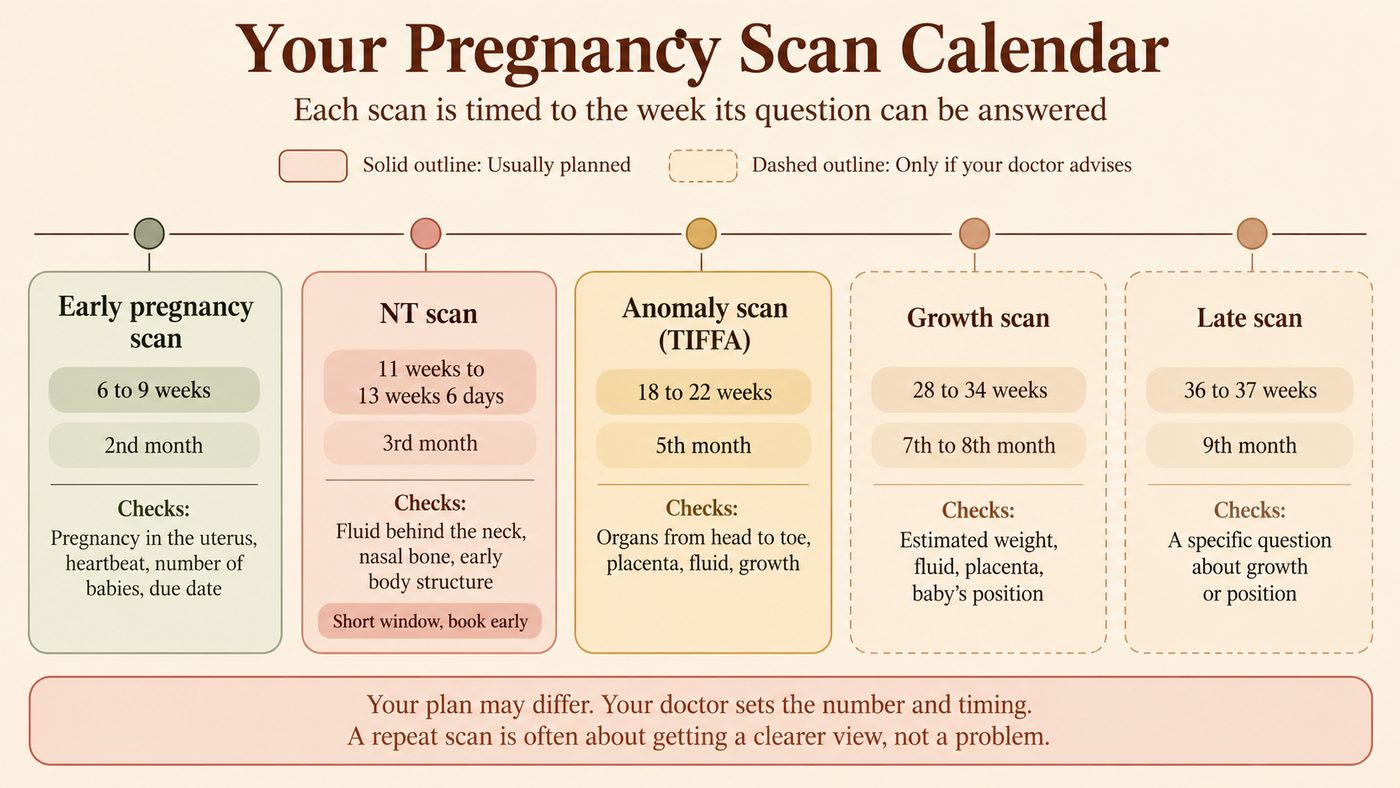
| Scan | When | Pregnancy month | What it checks | How routine is it? |
|---|---|---|---|---|
| Early pregnancy (viability) scan | 6 to 9 weeks | 2nd month | Pregnancy is in the uterus, heartbeat, number of babies, due date | Commonly done in India; timing varies |
| NT scan | 11 weeks to 13 weeks 6 days | 3rd month | Fluid behind the neck, nasal bone, early body structure, due date | Widely offered as screening |
| Anomaly scan (TIFFA or level 2) | 18 to 22 weeks | 5th month | Organs from head to toe, placenta, fluid, growth | Recommended in most guidelines |
| Growth scan | 28 to 34 weeks | 7th to 8th month | Estimated weight, fluid, placenta, baby's position | Offered by many Indian doctors; not routine in all guidelines |
| Late scan | 36 to 37 weeks | 9th month | A specific question about growth or position | Only if advised for a reason |
The first scan confirms the pregnancy and the heartbeat
Your first ultrasound is usually done between 6 and 9 weeks. It is often called the early pregnancy or viability scan. It confirms the pregnancy is in the uterus, checks the heartbeat and counts how many babies there are.
Around 6 weeks, your baby is only a few millimetres long, about the size of a lentil. Yet the tiny flicker of a beating heart can often already be seen on the screen.
Because your baby is so small, the scan may be done internally with a thin probe (transvaginal ultrasound). Whether the heartbeat is seen depends on exactly how far along you are, the type of scan and individual factors. Not seeing it yet does not, by itself, mean something is wrong. Your doctor will decide whether and when to repeat the scan.
Why does the NT scan have such a short window?
The NT scan can only be done between 11 weeks and 13 weeks 6 days. The fluid behind your baby's neck, called nuchal translucency, can be measured reliably only while your baby is within a certain size range.
On its own, the NT measurement is not a diagnosis. In many clinics it is combined with the double marker blood test and your age to give an estimate, such as a 1 in 1,000 or a 1 in 100 chance of conditions like Down syndrome. Tests and calculations vary between centres.
A "higher chance" result means further tests are offered, not that your baby has the condition. In the United Kingdom's national programme, for example, most people with a higher-chance result have a baby without these conditions. Figures in India depend on the test used, so ask your doctor what your own number means.
The anomaly scan is the most detailed look at your baby
The anomaly scan is done between 18 and 22 weeks, in the 5th month. It is also called TIFFA, short for Targeted Imaging for Fetal Anomalies, or a level 2 scan. It usually takes 30 to 45 minutes and checks:
- the brain, face and spine
- the heart and its chambers
- the kidneys, stomach and limbs
- your baby's growth, the placenta and the amniotic fluid
Your report will usually fall into one of these groups:
| What the report shows | What it usually means |
|---|---|
| All structures seen and normal | Very reassuring for the conditions the scan can see |
| Incomplete view | A part could not be seen clearly, often because of your baby's position; a repeat view is arranged |
| Soft marker | A small feature whose meaning depends on which marker it is and on your other screening results; many turn out to be normal variations |
| Finding that needs assessment | The next step depends on what was seen and may include a specialist scan or other tests |
A normal report is genuinely good news. Still, some conditions are too subtle to see, develop later, or cannot be seen on ultrasound at all. No scan can promise that every condition has been ruled out.
It is common to walk in excited and walk out anxious over one unfamiliar word on the report. A soft marker describes something the scan saw. It does not mean your baby has a condition, and your doctor will explain whether it matters in your case.
Is a third-trimester growth scan always needed?
Not always. Many doctors in India offer one between 28 and 34 weeks, but international guidelines do not recommend it routinely for every low-risk pregnancy. Your doctor may suggest it if your bump measures smaller or larger than expected, or if you have high blood pressure, diabetes or a placenta concern.
The scan usually checks:
- your baby's estimated weight
- the amount of amniotic fluid
- where the placenta lies and which way your baby is facing
Estimated weight is calculated from measurements of the head, tummy and thigh bone, so it is not an exact weight. The margin varies with the method, the stage of pregnancy and your baby's size. One reading is a snapshot. Two readings a few weeks apart show whether your baby is following a steady curve, which is usually what matters most.
A Doppler study, which measures blood flow through the umbilical cord, is usually added when there is a specific reason, such as slow growth or high blood pressure. A late scan around 36 to 37 weeks is not routine. It is done only for a particular question, such as your baby's position or growth.
What if a scan has to be repeated?
Being asked to come back is common. It usually means the picture was incomplete, not that something worrying was seen. The reason decides how soon you return.
| Reason | What usually happens |
|---|---|
| Your baby's position blocked a view | A short walk or snack, then a second try, or a new appointment in a few days |
| Too early to see something clearly | A repeat scan when your baby is a little bigger, timed by your doctor |
| Images were unclear | Another appointment, or an internal scan for a closer view |
| A measurement sits near a cut-off | A repeat to see the trend rather than rely on one number |
| A soft marker or finding | A detailed scan, often with a fetal medicine specialist |
Some pregnancies need extra scans
Some health situations change what your doctor needs to watch. A series of scans shows a trend that a single scan cannot. The schedule is set for each pregnancy individually, so two people in the same situation may have different plans.
| Situation | Why your doctor may recommend additional monitoring |
|---|---|
| Twins or more | To track each baby's growth and how the placenta is shared |
| Pregnancy through IVF (in vitro fertilisation) | Sometimes an earlier scan to confirm location and heartbeat; later care is often routine |
| High blood pressure or diabetes | Depending on control and severity, to follow growth, fluid and blood flow |
| Bleeding or a low-lying placenta | To see whether the placenta moves up as the uterus grows |
| Baby measuring small or large | To check whether growth follows a steady curve |
| Previous pregnancy loss or preterm birth | Depends on the cause; some need cervix checks, many need no extra scans |
How safe are repeated ultrasounds?
Ultrasound is considered safe when it is done for a medical reason by trained staff. It builds a picture from echoes of sound too high for you to hear. Unlike X-rays, it does not use radiation that can damage cells.
A long-term study followed children up to 8 years of age. It found no differences in the speech, language, behaviour and neurological development it measured after repeated scans. No single study covers every situation, and energy use varies by scan type, with Doppler using more. That is why experts advise using ultrasound only when it answers a medical question, and for the shortest time needed.
For the same reason, the United States Food and Drug Administration advises against "keepsake" sessions done only for photos or videos. They add exposure without checking your baby's health.
A little preparation helps
- Carry all previous scan and blood reports.
- Follow the scan centre's instructions on your bladder and food.
- Wear a loose two-piece outfit so your tummy is easy to reach.
- Carry your Mother and Child Protection card or pregnancy file, if you have one.
- Ask when your doctor will explain the report, especially if a word on it is unfamiliar.
Looking ahead
Once the anomaly scan is done, scans stop being your only window. From around 28 weeks, you will come to know your baby's usual pattern of kicks and rolls. Any change in that pattern, especially fewer movements, should be reported to your doctor the same day, without waiting for the next scan.
The final scan shifts the question from "Is my baby developing well?" to "How will my baby be born?" Position can still change in the last weeks, and weight is only an estimate. Your doctor combines the scan with an examination before planning the birth.
When to speak to a doctor
Some signs need care straight away. Others can wait for a call or your next visit.
Seek care urgently (go to your hospital or call your doctor now) if you have:
- heavy bleeding at any stage
- severe or one-sided tummy pain in early pregnancy, especially with dizziness or shoulder pain
- leaking fluid from the vagina
- fewer baby movements than usual after 28 weeks
Contact your doctor soon if:
- you have light spotting that does not settle
- you have crossed 13 weeks without an NT scan
- no anomaly scan has been planned by 20 weeks
- a word on your scan report worries you or is unclear
References
- World Health Organization. WHO recommendations on antenatal care for a positive pregnancy experience. 2016.
- World Health Organization. New guidelines on antenatal care for a positive pregnancy experience (news release). 2016.
- World Health Organization. Maternal and fetal assessment update: imaging ultrasound before 24 weeks of pregnancy. 2022.
- Salomon LJ, Alfirevic Z, Berghella V, et al. ISUOG Practice Guidelines (updated): performance of the routine mid-trimester fetal ultrasound scan. Ultrasound Obstet Gynecol. 2022;59(6):840 to 856.
- International Society of Ultrasound in Obstetrics and Gynecology. Practice Guidelines, including Performance of 11 to 14 week ultrasound scan (2023) and Performance of third-trimester obstetric ultrasound scan (2024).
- National Health Service (UK). Screening for Down's syndrome, Edwards' syndrome and Patau's syndrome.
- National Health Service (UK). 20-week screening scan.
- Newnham JP, Doherty DA, Kendall GE, et al. Effects of repeated prenatal ultrasound examinations on childhood outcome up to 8 years of age: follow-up of a randomised controlled trial. Lancet. 2004;364(9450):2038 to 2044.
- United States Food and Drug Administration. Avoid Fetal "Keepsake" Images, Heartbeat Monitors (Consumer Update).
- Government of India. The Pre-conception and Pre-natal Diagnostic Techniques (Prohibition of Sex Selection) Act, 1994. India Code.
Common questions
How many ultrasounds are normal during pregnancy?
The World Health Organization recommends at least one scan before 24 weeks. In India, many doctors plan three to four: an early scan, the NT scan, the anomaly scan and sometimes a growth scan. More may be added for twins, diabetes, high blood pressure or a placenta concern. The number matters less than the reason. Each scan should answer a specific question about you or your baby.
Can the ultrasound tell me if my baby is a boy or a girl?
Not in India. Under the Pre-Conception and Pre-Natal Diagnostic Techniques (PCPNDT) Act, it is illegal for any clinic or doctor to tell you your baby's sex, whether by words, signs or any other method. Scan centres must follow this strictly, so it is best not to ask. The scan's purpose is your baby's health and development.
Do I need a full bladder for a pregnancy scan?
It depends on the scan. For an internal (transvaginal) scan in early pregnancy, you are usually asked to empty your bladder. For some scans over the tummy in the first trimester, a moderately full bladder gives a clearer view. Later scans usually need no special preparation, so check with your scan centre when you book.
Is a 3D or 4D scan better than a normal scan?
Not for medical purposes. The standard two-dimensional scan is what doctors rely on to check your baby's organs and growth. A 3D (three-dimensional) or 4D (four-dimensional, moving) view shows the baby's surface, such as the face, and is sometimes used to look more closely at a specific finding. If you want one, ask for it as part of a medically advised scan rather than a separate photo session.
What happens if I miss the NT scan window?
After 14 weeks, the neck fluid can no longer be measured reliably, so the NT scan cannot be done. Depending on how many weeks you are and any tests already done, your doctor may suggest the quadruple marker blood test (usually 15 to 20 weeks) or NIPT (non-invasive prenatal testing), a blood test. These screen in different ways and do not give exactly the same information as the NT scan. The anomaly scan still checks your baby's body in detail.
What does it mean if my baby is measuring smaller or larger than expected?
Babies come in different sizes, and one measurement slightly above or below average is common. It often reflects family build or small differences between measurements. Doctors look at your baby's percentile and, more importantly, whether growth follows a steady curve over time. If growth slows or is well outside the usual range, your doctor may suggest a Doppler study and more frequent scans.
Can I ask for an extra scan between routine appointments?
You can always discuss it with your doctor. If you have bleeding, pain, leaking fluid or fewer movements, seek care promptly; a scan may be part of that check. Scans purely for reassurance are not usually recommended. If worry between scans feels heavy, tell your doctor, who may listen to the heartbeat or talk through what you are feeling.
This article is for general education and helps you understand how pregnancy ultrasounds are usually planned. It is not a substitute for personal medical advice. Scan timing can differ based on your health and your pregnancy, so please follow the schedule your doctor recommends and discuss any concerns with them.
 Medically reviewed byDr. Mahender Singh, MBBS, DCH, MD (Paediatrics)A consultant child specialist and neonatologist with over four decades of practice in newborn and child health.
 Written bySarthak ThakurCo-founder, CTO and Chief Marketing Officer of ParentVeda, and the software engineer who builds its app and the pipeline that checks its articles.
This article explains; it doesn’t diagnose. If your doctor has told you something different, your doctor is right.